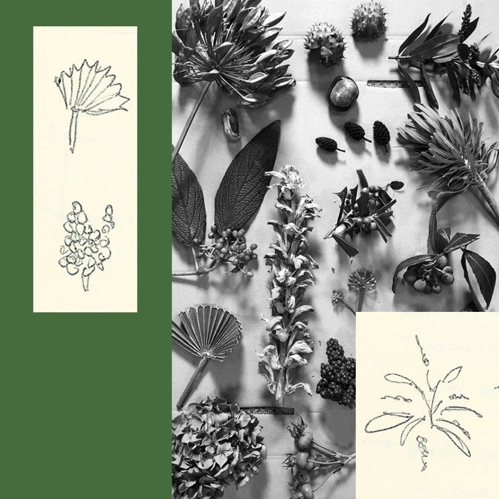
Sketchbook_
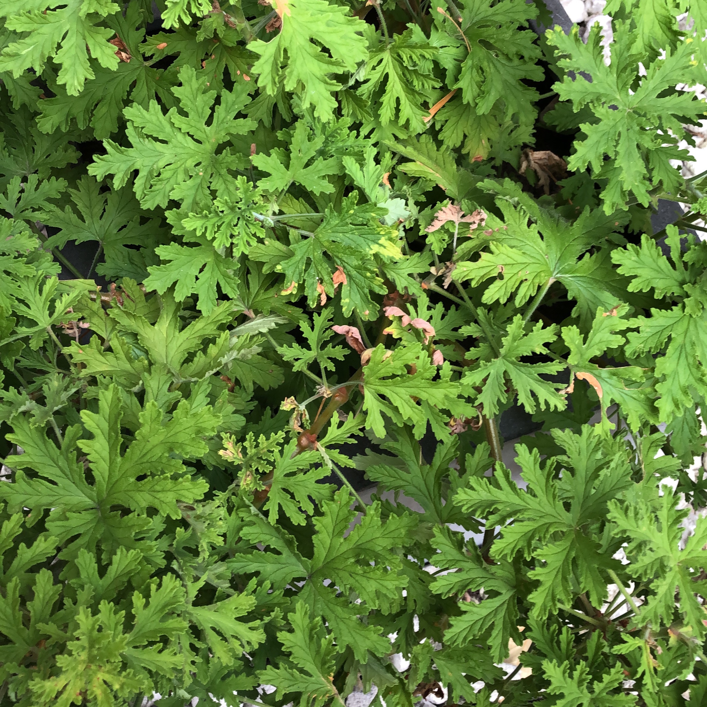
Thumbnail sketches and samples of plant life collected around the hospital campus – berries, buds, seedpods etc. for use in the scanners.
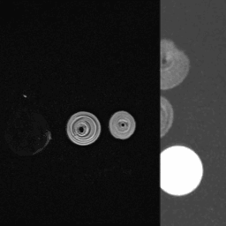
After the initial MRi scanning and consultation with radiographers I aimed to find denser items which would be more likely to be detected in the scanners.

The roof tiles above CT and MRI scanners had been proposed areas to host my outcomes. During my visit I was interested to see these and what imagery is currently in place.
After consultation with imaging staff, placing work here may offer a tangible effect on the patient experience, creating a conversation point to calm patients before scans as well as a level of escapism and alternative interest in a stressful environment.
Definitely an area to explore and find out technical possibilities.

MRi Honey Locust animation_
Sequenced scans of Honey Locust branches. I’m really enjoying the ethereal quality of these scans and the potential narratives which emerge when the scans are sequenced together. Massive thanks to Chief MRi Radiographer Rick for capturing these images and enabling my strange requests!
These were the first plant samples I found, by chance in the morning on the first day of the residency. I noticed the tree being trimmed on Sydney Street and asked the tree surgeon if I could take some. Apparently it’s quite strange to find Honey Locust in this area.

Thinking more about King’s Garden, the idea of small, secret garden spaces was a recurring theme throughout my visit. The King’s Garden is a quiet space at the back of the Fulham Wing used for physiotherapy and containing ‘sensory’ plants, as I learnt from the gardener Carl.

CT Scanning test and a secret garden wall_
As well as the MRi scanning, on the first day I was able to test CT scanning with Radiographer Aphoung. Here are some of the plants I had collected from the hospital campus on the first day (Zamias and Honey Locust branch) going into the scanner. This is collaged with a grid pattern I noticed in the King’s Garden. The idea of grids/geometric structure juxtaposed with natural forms was something I was drawn to throughout my visit.

MRI Bromelia animation_
The sequential nature of the scanning lends itself to moving image. This animation test is created using axial MR scans of a Bromelia plant.
The images from the scanner come out at a relatively low resolution, but I am interested in the distortion and noise this creates when the image is presented at a larger size. How images are made is a key interest, especially given the context of an Imaging Centre.

Research MRI scanning_
A key part of the residency is to explore medical diagnostic scanning equipment as image making tools, focusing on plant life found around the area. This collage features imagery of a Bromelia plant I found on the hospital campus created by an MRI scanner, with the expertise and help of Chief MRi Radiographer Rick.

Day 1_
My first visit was between 14-19th September. My plan was to begin to explore the area to inform my eventual outcomes work. I spent the first day getting my bearings and noting anything of immediate interest.

Design a site like this with WordPress.com
Get started